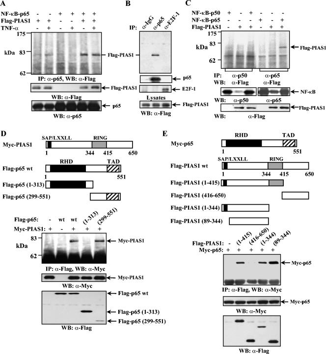
FIG. 1.
PIAS1 specifically interacts with the p65 subunit of NF-κB in vivo. (A, top panel) Human 293T cells were transiently transfected with either Flag-PIAS1 or p65 alone or together, as indicated. Thirty hours posttransfection, cells were either untreated or treated with TNF-α for 15 min. The whole-cell lysates were subjected to coimmunoprecipitation (IP) with anti-p65 (Santa Cruz Biotechnology) followed by Western blotting (WB) with anti-Flag (Sigma). (Middle and bottom panels) The same lysates were subjected to sodium dodecyl sulfate-polyacrylamide gel electrophoresis and probed with anti-Flag or anti-p65 as indicated. (B) The same procedure described for panel A was performed except that 293T cells were transiently transfected with both Flag-PIAS1 and NF-κB p65, and coimmunoprecipitation assays were performed with rabbit IgG, anti-p65, or anti-E2F-1 (Santa Cruz Biotechnology) followed by Western blotting with anti-Flag (top panel). The same filter was reprobed with anti-p65 or anti-E2F-1 (middle two panels). The same lysates were also analyzed by Western blotting with anti-Flag to show equal amounts of Flag-PIAS1 present in each sample (bottom). (C) The same procedure described for panel A was performed except that 293T cells were transfected with either Flag-PIAS1, p65, or p50 alone or together, as indicated, and coimmunoprecipitation assays were performed with anti-p50 (lanes 1 to 3) or anti-p65 (lanes 4 to 6) (top). The same lysates were probed with anti-p50, anti-p65, or anti-Flag, as indicated (middle and bottom panels). (D) The same procedure described for panel A was performed except that Myc-PIAS1 and Flag-p65 wt, Flag-p65(1-313), or Flag-p65(299-551) was used as indicated. (E) The same procedure described for panel A was performed except that Myc-p65 and Flag-PIAS1 wt or different mutants were used as indicated.

