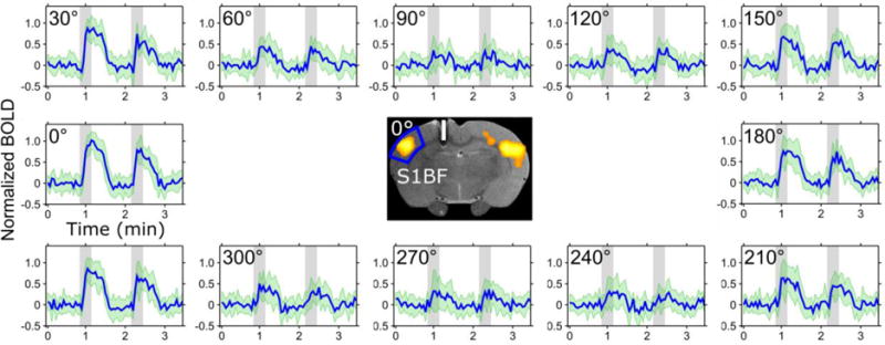
Figure 3.

Activation resulting from directionally selective biphasic pulses applied in the CC in the rat brain. Time series averaged over 12 animals for different orientations of the electric field relatively to the axonal track in the CC. When the principal direction of the dipolar field was orientated at 90° and 270° relative to the axonal track, no BOLD contrast was observed during the stimulation. The ROI for the time series is shown in the middle with a representative BOLD activation map at 0° overlaid on a FSE image where the white bar represents an estimate of the electrode dimensions and placement in the brain based on Paxinos’ rat atlas [16]. The SDs are indicated in green.
