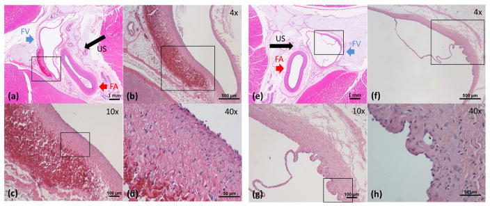
Figure 8.
Histology of Femoral Veins. The direction of ultrasound propagation is indicated by black arrow. (a)–(d): Femoral vein wall right after a free-flow treatment. In (a), the lumen with thin vessel wall on the left is the femoral vein (FV, blue arrow) and the lumen with thick wall on the right is the femoral artery (FA, red arrow). The tissue that immediately surrounds the vessels is connective tissue and the dense, outer surrounding is skeletal muscle. (b)–(d) are magnified images of the area in the box in the previous image. There was focal venular wall thickening with corresponding hemorrhage in the tunica adventitia and thickening of the tunica media. Most of the tunica intima of the femoral vein was intact. No thrombus was noted in the lumen. No injury was found in or around the femoral arteries. (e)–(h): Femoral vein wall two weeks after a thrombolysis treatment. (f)–(h) are magnified images of the area in the box in the previous image. There was a mild thickening of the tunica media with granulation tissue composed of small vascular channels and inflammatory cells. There was no hemorrhage or evidence of tunica intima injury. No injury to the femoral arteries and surrounding tissues as well. Valves in the femoral veins were preserved.

