
Fig. 1.
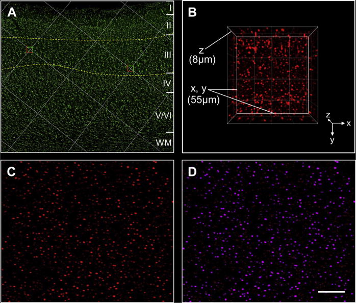
Image collection and processing. (A) A section of PreC processed using fluorescent Nissl histology illustrates cortical laminae I to VI, delineation of lamina III, and selection of sampling sites at grid intersections within the outlined area; (B) a 3-dimensional view of an image stack of spinophilin immunofluorescence in PreC lamina III with X, Y, and Z axes indicated; (C) a single-plane image showing spinophilin-ir puncta after camera background subtraction, deconvolution, and Gaussian filter transformation; (D) examples of masked (selected) spinophilin-ir puncta (purple). Each selected puncta is an “object” with measured attributes such as intensity obtained using Slidebook software. Scale bar = 400 μm for A and 20 μm for C and D. Abbreviation: PreC, precuneus.