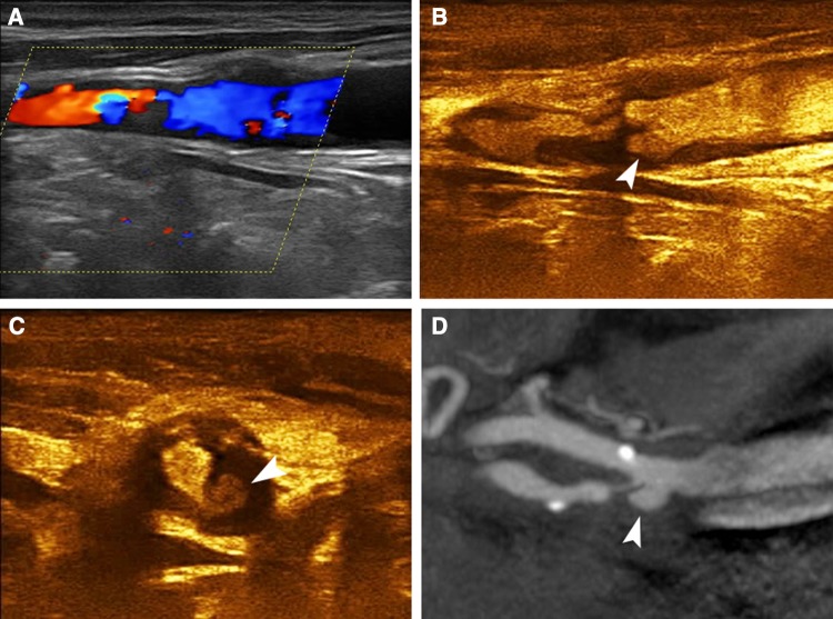
Fig. 2.
A 77-year-old asymptomatic male patient with ulcerated carotid plaque. Colour Doppler imaging a identifying a highly stenotic plaque with relatively irregular surface, which however could not be accurately delineated. Long-axis CEUS image b revealing the presence of a type 3 ulcer (arrowhead). Short-axis CEUS image c confirming the ulcer’s presence (arrowhead). MDCTA d confirmed the presence of a highly stenotic and ulcerated carotid plaque (arrowhead)

