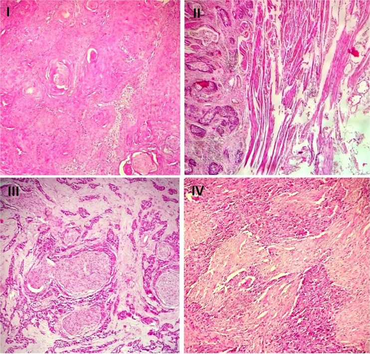
Fig. 2.
Histological pictures of four ESCC cases with different stages. I. A well differentiated squamous cell carcinoma with keratin pearl formation and minimal nuclear atypia. A stage I tumor with GLI1 overexpression (H&E stain, ×200). II. The moderately differentiated tumor invades muscularis propria with stage II. GLI1 and SOX2 overexpression was detected in this sample (H&E stain, ×100). III. The poorly differentiated tumor with perineural invasion in ESCC. A stage III tumor with GLI1 and SOX2 overexpression (H&E stain, ×200). IV. The poorly differentiated squamous cell carcinoma demonstrates pleomorphic cells with high nucleo-cytoplasmic ratio and elevated expression of SOX2. The patient had distant metastasis (stage IV) (H&E stain, ×200)

