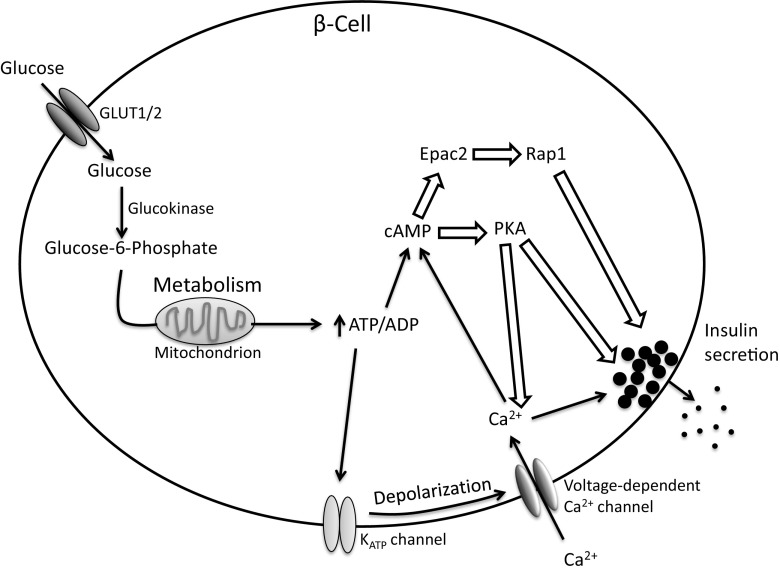
Fig. 2.
Glucose-stimulated insulin secretion pathway. Glucose enters the β-cell via GLUT2 (mouse) or GLUT1 (human) where it is phosphorylated to glucose-6-phosphate by glucokinase. Glucose-6-phosphate is metabolized in the mitochondria, generating ATP. An increase in the ATP/ADP ratio leads to closure of KATP channels, membrane depolarization, and subsequent opening of voltage-dependent Ca2+ channels. Increased cytosolic Ca2+ levels triggers insulin granule release. ATP can also lead to formation of cAMP. cAMP signals via protein kinase A (PKA) or exchange factor directly activated by cAMP (Epac) 2 and can amplify insulin secretion. Hollow block arrows represent components also regulated by PGs that may be involved in altering insulin release

