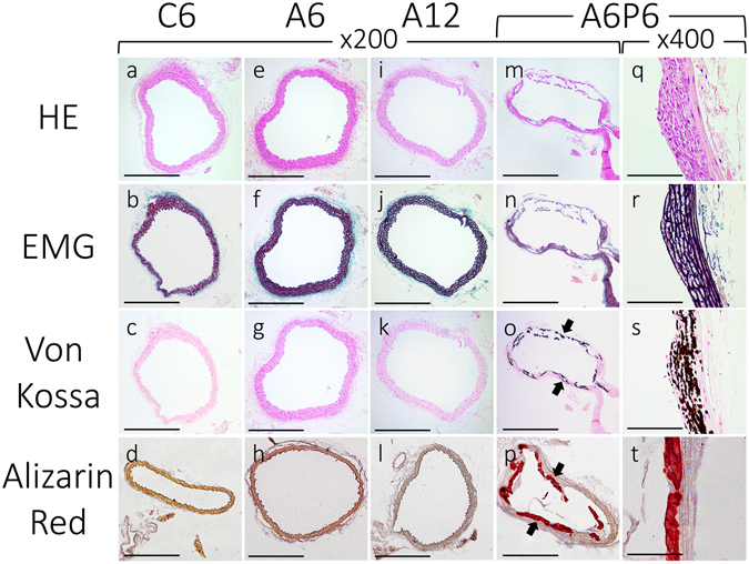
Figure 3.

Representative micrographs of H&E, EMG, Von Kossa and Alizarin Red stained sections of the thoracic aorta. Sections of the thoracic aorta for the control (a–d), A6 (e–h), A12 (i–l), and A6P6 (m–t) groups are shown. Von Kossa and Alizarin Red staining positive legions were observed in CKD-HP mice (o,p,s,t, black arrows). Images for the A6P6 mice are shown as representatives of all CKD-HP group, as calcification in A6P2 and A6P4 groups showed similar histological features (see Supplementary Fig. S1). Scale bar, 500 μm for (a–p) and 50 μm for (q–t); H&E, hematoxylin and eosin staining; EMG, elastica Masson-Goldner staining.
