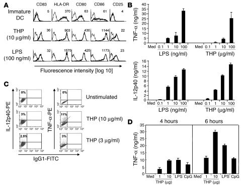
Figure 1.
THP activates professional APCs. (A) Immature monocyte-derived human DCs were stimulated with THP or LPS. Profiles with fine lines represent staining patterns with an isotype-matched control Ab, and profiles with bold lines represent staining with mAb of the indicated specificity. Data are representative of 5 independent experiments. Numbers indicate mean fluorescence intensity of specific Ab staining. (B) Immature human DCs were stimulated with different concentrations of THP or LPS. Cell-free supernatants were collected 18 hours after addition of THP or LPS and were analyzed by ELISA. Med, medium control. (C) Intracellular cytokines were stained in human DCs 18 hours after stimulation and then analyzed by FACS. Results are representative of 3 independent experiments. (D) Mouse RAW264.7 macrophages were exposed to THP, LPS, and CpG for the indicated time periods. Cell-free supernatants were analyzed for TNF-α by ELISA. Similar results were obtained in 3 other independent experiments and data are expressed as means ± SD of triplicate cultures in a representative experiment.

