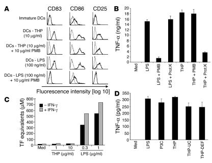
Figure 2.
DC maturation induced by THP is not due to LPS or protein contamination. (A) THP and LPS were left untreated or pretreated with polymyxin B (PMB) for 2 hours and then added to human immature DCs. After 48 hours, cells were harvested and analyzed by FACS. Profiles with fine lines represent staining patterns with an isotype-matched control Ab, and profiles with bold lines represent staining with a mAb of the indicated specificity. Data are representative of 5 independent experiments. (B) THP was incubated with PMB beads overnight. The supernatant was analyzed for the presence of THP by SDS-PAGE. Additionally, THP was incubated with proteinase K (Prot.K) for 45 minutes as described (11). The PMB-purified and the proteinase-treated samples were incubated with RAW 264.7 macrophages for 18 hours and analyzed for TNF-α. LPS as a control was treated similarly. Similar results were obtained in another independent experiment. (C) Effect of THP and LPS on the induction of TF activity in HUVECs. HUVECs were pretreated with or without IFN-γ and then exposed to THP or LPS. A 1-stage clotting assay was used to determine TF activity. The results are representative of 3 independent experiments. (D) LPS, Pam3Cys (P3C), THP isolated by standard NaCl precipitation (THP), THP isolated by NaCl precipitation and ultracentrifugation (THP-UC), and THP isolated by NaCl precipitation, ultracentrifugation, and diatomaceous earth filter (THP-DEF) were added to C57BL/6 splenocytes for 20 hours. Cell-free supernatants were analyzed for TNF-α by ELISA.

