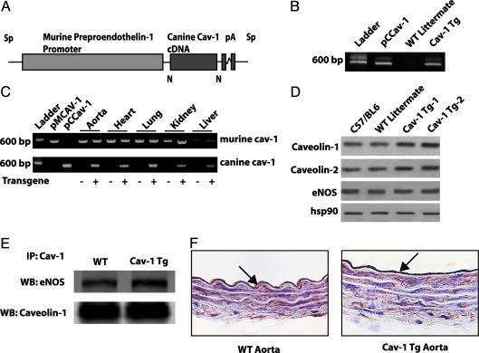
Fig. 1.
Generation of endothelium-specific TG mice. (A) Construct used for generating the Cav-1 TG mice. The canine Cav-1 cDNA was driven by the preproendothelin promoter. (B) Expression of the transgene vs. endogenous Cav-1. Primers specific for canine Cav-1 recognize the plasmid containing canine Cav-1 (pCCav-1) and TG Cav-1 (Cav-1 TG) but not endogenous Cav-1 (WT littermate). (C) The presence of both WT and transgene Cav-1 by RT-PCR in various tissues. pMCav-1 is the murine Cav-1 cDNA (D) The increased expression of Cav-1 and Cav-2 in aortic extracts from two TG lines (TG-1 and TG-2), compared with C57Bl6 or WT littermates. (E) Immunoprecipitation of Cav-1 from TG lung extracts increases the amount of eNOS co-associated (Lower, total Cav-1 immunoprecipitated; Upper, coassociated eNOS). (F) Immunohistochemical evidence for expression of Cav-1 transgene in the endothelium. WT and Cav-1 TG aortic sections were stained with a Cav-1 mAb. Note the presence of intense labeling of Cav-1 in the EC, with little difference in the intensity of Cav-1 in underlying smooth muscle.

