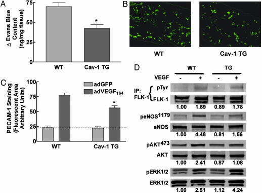
Fig. 4.
VEGF-mediated functions are reduced in Cav-1 TG mice. (A) Cav-1 TG mice exhibit reduced (30 min) VEGF-stimulated vascular leakage. Data are mean ± SEM (n = 6). (B) PECAM-1-positive vascular structures in the ears of WT (Left) or Cav-1 TG (Right) mice injected with AdVEGF (after 6 days). (C) AdVEGF-mediated angiogenesis was reduced in Cav-1 TG (mean ± SEM, with n = 6 animals per group, and 10 fields were quantified for each ear). (D) VEGF-mediated signal transduction to Akt and eNOS are reduced in Cav-1 TG mice. Mice were injected with saline or VEGF, and lung tissue was processed for Western blot analysis. The numbers below represent densitometric evaluation of the data, with a value of 1.0 reflecting the basal level of phosphorylation of each signaling protein (FLK, eNOS, Akt, and ERK, respectively). Similar results were obtained in three additional experiments. *, P < 0.05.

