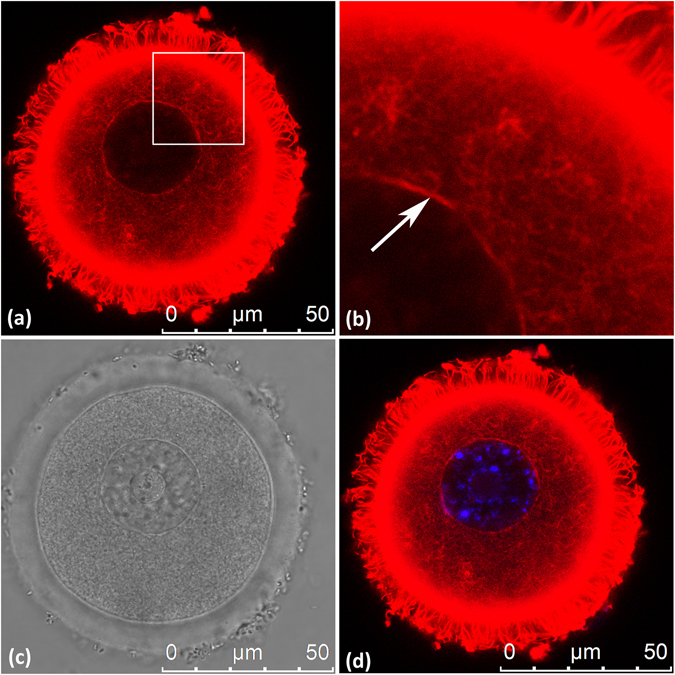
Figure 3.

Nuclear actin rim surrounds the GV of mouse oocytes. GV oocytes were fixed and processed as described in ‘Experimental Procedures’. Actin filaments were stained with Tritc-Phalloidin (red) diluted in PBS, containing 0.1% Triton X-100 and 3% BSA. DNA was stained with Hoecst (blue). (a) Tritc-Phalloidin; (b) Enlargement of the area marked in (a); (c) Bright field; (d) Merge of Tritc-Phalloidin and Hoecst. White arrow points at the actin rim.
