
Figure 3.
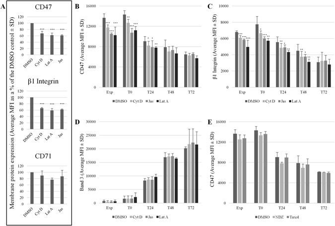
CD47 membrane stability is reduced following incubation with actin disrupting drugs. (A) K562 cells or (B–D) expanding (Exp) and differentiating (T0-T72) erythroblasts were incubated for 30 minutes with Cytochalasin (Cyt) D (5 µM), Latrunculin (Lat) A (1 µM), Jasplakinolide (Jas; 1 µM) or a DMSO control. (E) Erythroblasts were incubated with the microtubule stabilising drug paclitaxel (Taxol; 10 µM), the microtubule destabilising drug nocodazole (NDZ; 10 µM), or a DMSO control for 3 hours (n = 2). Treated cells were then incubated with BRIC32 (anti-CD47) and P5D2 (anti-β1 integrin). (A) CD71 (anti-Transferrin receptor) was used as a negative control in K562 cells (average MFI as % of the DMSO control ± SD (n = 3); ***p ≤ 0.001, **p ≤ 0.01, *p ≤ 0.05 using the Students T test). BRIC71 (anti-band 3) was used as a negative control in erythroblasts (average MFI ± SD (n = 3)). Cells were then incubated with an APC-conjugated monoclonal IgG1 anti-mouse secondary antibody. Emissions from 10,000 events were detected using a MACSQuant Analyser 10 flow cytometer and data was analysed using FlowJo version 10. The MFI were shown in (B-D) due to the differences in surface expression known to occur during terminal differentiation.