Fig. 2.
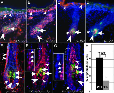
Expression patterns for BMP receptors and pSmad1/5 in developing and cycling HFs of WT and K5-Noggin mice. Cryosections of the dorsal skin of WT and TG mice harvested at P2.5 (A–D) or 5 days after depilation (E–H) were processed for double immunovisualization of BMPR-IA, BMPR-IB, pSmad1/5 (red fluorescence), and Trp-2 (green fluorescence). Cryosections were counterstained by DAPI (blue fluorescence). (A and B) BMPR-IA expression in the epidermis (small arrows), hair bulb (arrowheads), and lack of expression in the follicular MCs (large arrows) of stage 5 HFs of WT (A) and TG (B) mice. (C and D) Decrease of pSmad1/5 expression in the epidermis (small arrowheads), dermal papilla (large arrowheads), and hair matrix (small arrows) of stage 5 HF in TG mice (D) vs. the corresponding compartments of a WT mouse (C). Lack of expression in the follicular MCs (large arrows) is shown. (E) BMPR-IA expression in the hair matrix (small arrows), outer root sheath (small arrowheads), MCs (large arrows), and dermal papilla (large arrowhead) of anagen IV HF in the skin of a WT mouse. (F and G) Decrease of nuclear pSmad1/5 expression in the outer root sheath and hair matrix (small arrows) of anagen IV HF in TG (G) compared with WT (F) mice. pSmad1/5 expression in the inner root sheath/hair shaft (small arrowheads), dermal papilla (large arrowheads), and Trp-2+ MCs (large arrows) is indicated. (Insets) High magnification of the labeled areas. (H) Histomorphometry of the pSmad1/5 positive cells in the outer root sheath of anagen IV HFs in TG and WT mice (mean ± SEM, **, P < 0.01, Student's t test).

