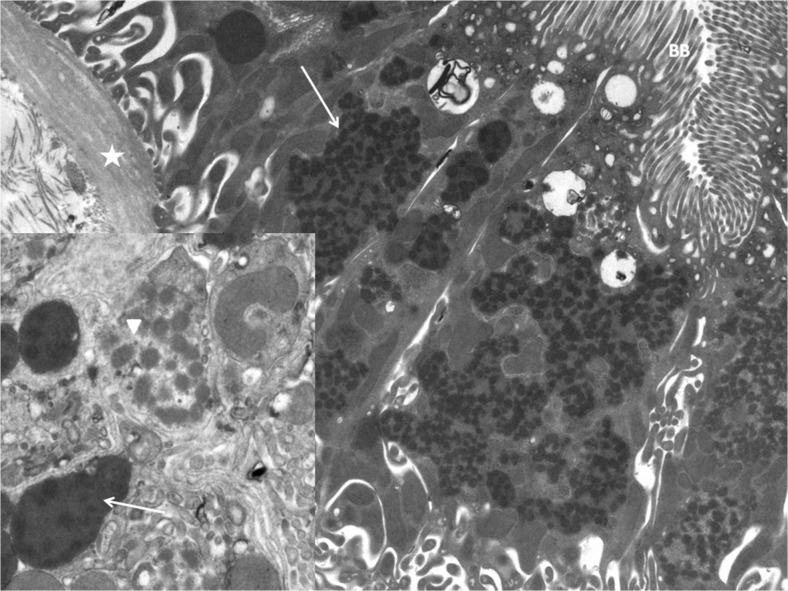
Fig. 1.
Electron microscopic image of kidney biopsy showing notable abnormal mitochondria. Asterisk Basal membrane, BB brush border proximal tubule, arrow fused mitochrondria. Inset: Arrowhead Swollen mitochondria exhibit electron-lucent areas, arrow fused mitochondria present amorphous matricial densities

