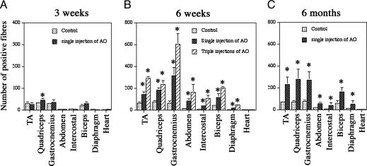
Fig. 2.
The number of positive fibers expressing membrane-localized dystrophin 2 weeks after the 2OMeAO treatments. mdx mice were treated at the age of 3 weeks (A), 6 weeks (B), and 6 months (C). Control, mdx injected with saline; TA, tibialis anterior; AO, 2OMeAO; Triple injections, injections at weekly intervals. The mean percentages of dystrophin-positive fibers in the 6-week-old group were 6%, 7.2%, 8.6%, and 8.2% in the AO-treated TA, quadriceps, gastrocnemius and biceps, respectively, in comparison with the 1.8%, 1.4%, 2.0%, and 1.7% in the control mice. The percentages were 12.5%, 9.4%, 16.2%, and 13.9% after triple injections. The percentages in the 6-month-old group were 8.9%, 11.4%, 7.8%, and 9.5% in the AO-treated TA, quadriceps, gastrocnemius, and biceps, respectively, in comparison with 2.8%, 3.1%, 2.6%, and 3.2% in the control mice. The percentage of dystrophin-positive fibers in the 3-week-old group was <1% in all four muscles except in the AO-treated quadriceps, which is 1.2%. The larger percentage of positive fibers when compared with the percentage of protein levels (Fig. 3A) may be attributed to the presence of revertant fibers, the lower-than-normal levels of the protein in most dystrophin-positive fibers, and the use of maximum numbers of dystrophin-positive fibers for each muscle sample. Sections were stained with polyclonal antibody P7 to dystrophin and detected with Alexa Fluor 594-labeled goat anti-rabbit Igs. (*, P < 0.05, ANOVA test; n = 4–6 mice).

