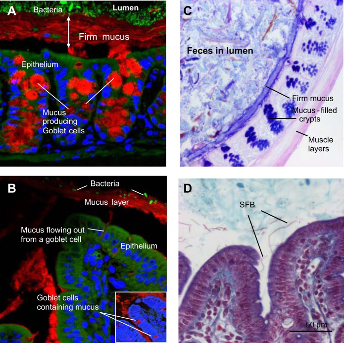
Fig. 2.
Fluorescence microscopy of mucus and microbiota in Carnoy-fixed sections of colon (A) and ileum (B) from mice. Mucin 2 (Muc2) was detected by immunofluorescence using anti-Muc2 and goat-anti-rabbit Alexa Cy3 antibodies (red). Nuclei were visualized using DRAQ5 (blue). Bacteria were identified using fluorescence in situ hybridization and the universal Euprobe 388 (green). C: Alcian blue/periodic acid Schiff-stained colonic tissue (frozen section) from a mouse showing a dark blue firm mucus layer, dark blue-stained goblet cells, and fecal material in the lumen. D: section of ileum (formalin fixed) from a conventional mouse stained with the Crossmon procedure. Arrows indicate segmented filamentous bacteria (SFB), which in contrast to other commensals, are typically found in contact with the epithelium.

