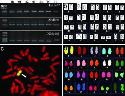
Fig. 3.
Genomic analysis of the sterile hermaphrodite mouse and its ntES cells. (a) PCR analysis of microsatellite markers in genomic DNA from the ear of the mouse (He), ntES cell lines (O1–O4), and a tetraploid complementation chimeric mouse (TC) confirms that they originated from the hermaphrodite mouse. The polymorphic markers D1Mit46, D2Mit102, and D4mit37 are conserved in genomic DNA from the hermaphrodite mouse, the ntES cell lines, and the tetraploid complementation chimeric mouse but differ from those of the oocyte recipient strain B6D2 F1 (F1). (b) Karyotype analysis from tail-tip fibroblast of tetraploid complementation chimera showed that the mice were made by diploid cells of ntES cell and hold the same Y chromosome abnormality as in donor ntES cells. (c) Fish chromosome painting of ntES cell with a mouse Y chromosome-specific probe. The probe hybridized to the metacentric region of the chromosome (arrow). Twenty-four of 25 metaphase (96%) showed this abnormality. (d) Spectral karyotyping FISH chromosome painting of ntES cell. There is no abnormality except for the Y chromosome.

