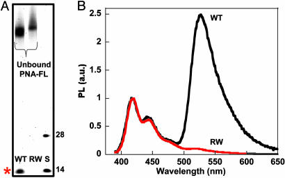
Fig. 6.
Fluorescence analysis of full-length ssDNA targets after enzymatic digestion. (A) PNA-FL was hybridized to 249-base WT and R406W sequences, and then digested with S1 nuclease. The 28- and 14-base size standards (S) correspond to the D0 sequence and the predicted 14-base product, respectively. The denaturing polyacrylamide gel was visualized via Sybr Gold staining (λex = 488 nm), and the band of interest is indicated by the red asterisk. (B) Normalized fluorescence of S1-treated hybridization products (WT, black; R406W, red). Spectra were obtained (λex = 380 nm) by adding CP directly to the quenched reaction mixtures diluted ([CP] = 1.4 × 10⁻⁷ M and [PNA/ssDNA] = 1 × 10⁻⁸ M) in 30 mM potassium phosphate buffer, pH 7.4.

