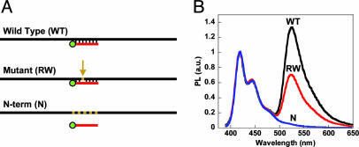
Fig. 1.
Fluorescence analysis of full-length ssDNA targets. (A) PNA-FL probe (red) annealed to asymmetric PCR products, with mutations indicated as tan segments in the black DNA sequence, for fluorescence analysis according to Scheme 1. (B) Normalized fluorescence of PNA-FL/ssDNA upon addition and excitation (λex = 380 nm) of the CP ([CP] = 4 × 10⁻⁷ M and [PNA/ssDNA] = 1 × 10⁻⁸ M) in 30 mM potassium phosphate buffer, pH 7.4, at room temperature. The black curve corresponds to the complementary (wild type) DNA, and the red and blue curves correspond to the single mismatch (RW) and noncomplementary (N-term) DNA targets, respectively.

