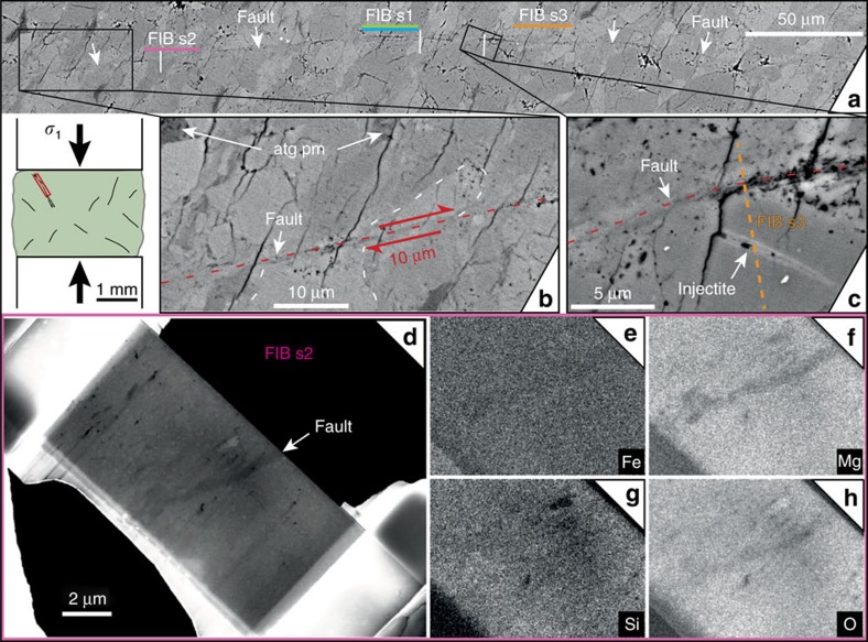
Figure 7. Microstructural observations in a sample with 5 vol% antigorite deformed at 1.1 GPa.
(a) Fault trace (complete in Fig. 6) with locations of FIB sections; (b,c) zooms into the fault: antigorite pseudomorphs (atg pm) and sheared grain (displacement ∼10 μm (b); position of FIB s3 (Fig. 8), crosscutting an injectite (c); (d–h) SEM on FIB section 2: density contrast (d) and EDX mapping (Fe, Mg, Si, O), showing that the fault is depleted in magnesium (e–h).

