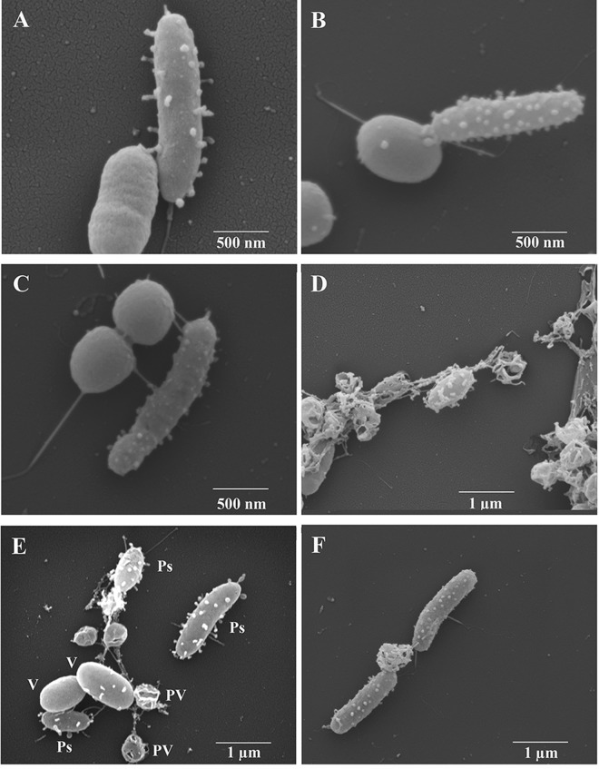
FIG 3.
Scanning electron micrographs of P. piscicida strains in coculture with V. parahaemolyticus O3:K6. (A to C) Apparent transfer of digestive vesicles from P. piscicida to the surfaces of V. parahaemolyticus in P. piscicida strain DE2-A (A and B) and strain DE1-A (C). (D) Vibrios containing vesicle-associated holes, except in center of micrograph, where an intact V. parahaemolyticus appears with vesicles on its surface. (E) P. piscicida (Ps), V. parahaemolyticus (V), and late-stage V. parahaemolyticus with vesicle-digested holes visible (permeabilized vibrios [PV]). (F) Two P. piscicida DE2-A bacteria suspected to be feeding (grazing) on nutrients released by a permeabilized V. parahaemolyticus.

