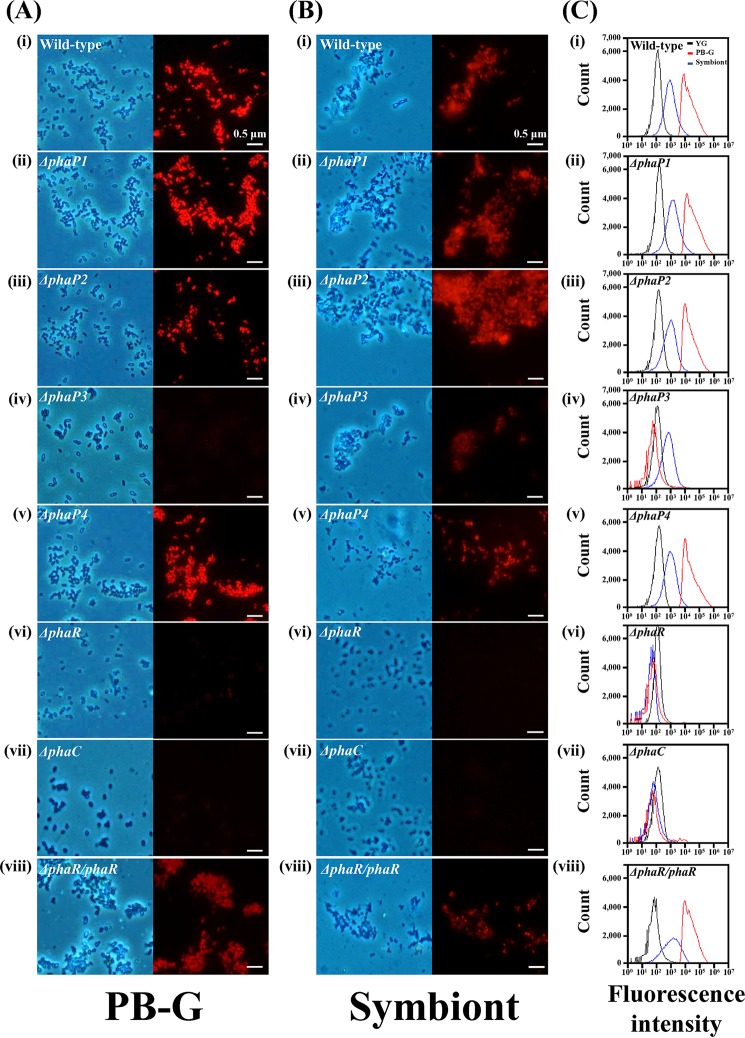
FIG 2.
In vitro PHA production by wild-type and PHA-related gene-depleted mutants of the Burkholderia symbiont. (A) Images of PHA-derived fluorescence under the fluorescence microscope. Phase-contrast images (left) and fluorescent images (right) of Burkholderia cells cultured in PB-G medium are stained with Nile blue A. (i) Wild type; (ii) ΔphaP1 mutant; (iii) ΔphaP2 mutant; (iv) ΔphaP3 mutant; (v) ΔphaP4 mutant; (vi) ΔphaR mutant; (vii) ΔphaC mutant; and (viii) ΔphaR/phaR Burkholderia mutant (scale bars, 0.5 μm). (B) Stained PHA granules of wild-type and mutant Burkholderia cells colonized in the midgut of fifth-instar nymph. (i) Wild type; (ii) ΔphaP1 mutant; (iii) ΔphaP2 mutant; (iv) ΔphaP3 mutant; (v) ΔphaP4 mutant; (vi) ΔphaR mutant; (vii) ΔphaC mutant; and (viii) ΔphaR/phaR Burkholderia mutant. (C) Flow cytometric histograms of PHA-derived fluorescence from Burkholderia cells cultured in YG medium (black), PB-G medium (red), and colonized symbiotic Burkholderia (blue). Burkholderia cells were stained with Nile blue A. (i) Wild type; (ii) ΔphaP1 mutant; (iii) ΔphaP2 mutant; (iv) ΔphaP3 mutant; (v) ΔphaP4 mutant; (vi) ΔphaR mutant; (vii) ΔphaC mutant; and (viii) ΔphaR/phaR Burkholderia strain.

