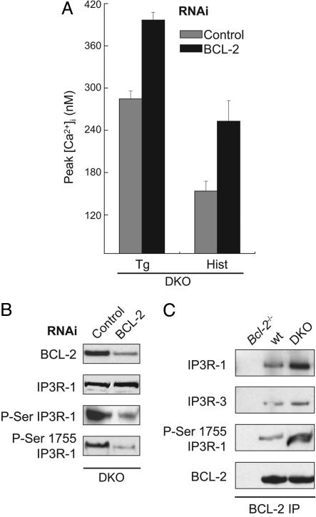
Fig. 2.
Knocking down BCL-2 expression reduces phosphorylation of IP3 receptor type 1 and increases Ca2+ release in DKO cells. (A) Peak intracellular Ca2+ concentration ([Ca2+]i) as measured by Fura-2 in response to thapsigargin (Tg) (200 nM) or histamine (Hist) (100 μM) in DKO mouse embryo fibroblasts (MEFs) transfected with control (gray bars) or Bcl-2 (black bars) RNAi 24 h before measurement. (B) Immunoblot of BCL-2 in total cellular lysates, or IP3R-1, phosphorylated serine (P-Ser) IP3R-1, and phosphorylated serine-1755 IP3R-1 in ER fractions of DKO MEFs 24 h after transfection with control or Bcl-2 RNAi. (C) BCL-2 immunoprecipitation from ER fractions of Bcl-2-/-, WT, and DKO MEFs. Immunoprecipitated complexes were then transferred to a membrane and blotted with antibodies against IP3R-1, IP3R-3, phosphorylated serine-1755 IP3R-1, and BCL-2.

