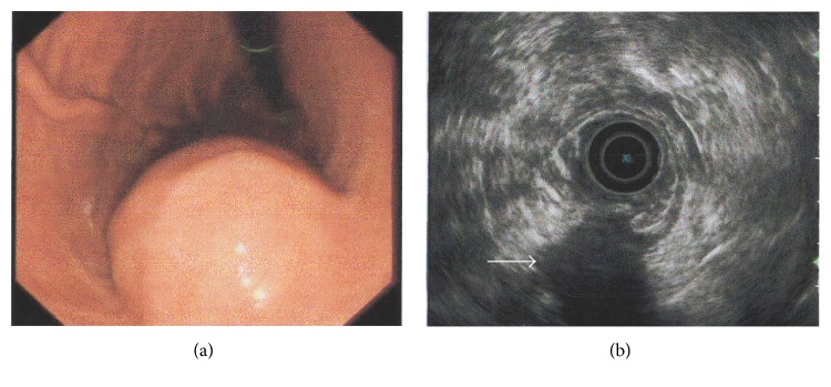
Figure 1.
Endoscopic (a) and endosonographic (b) findings in the current case of gastric schwannoma. (a) A round, submucosal mass with an indistinct border was observed at the lesser curvature of the gastric body. (b) On endoscopic ultrasonography, the lesion (white arrow) appeared homogeneous and its echogenicity was lower than that of the normal muscle layer. The mass measured 3.7 × 3.2 cm and originated from the fourth layer.

