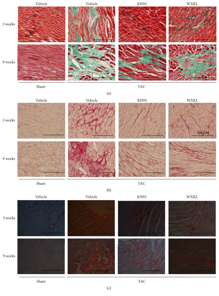
Figure 4.
Representative images of (a) Masson's trichrome staining method to evaluate cardiac fibrosis of each group at 3 and 9 weeks (n = 5), (b) Sirius Red staining to evaluate cardiac fibrosis of each group at 3 and 9 weeks (n = 5), and (c) Sirius Red staining to evaluate cardiac fibrosis of each group at 3 and 9 weeks under polarized light (n = 5).

