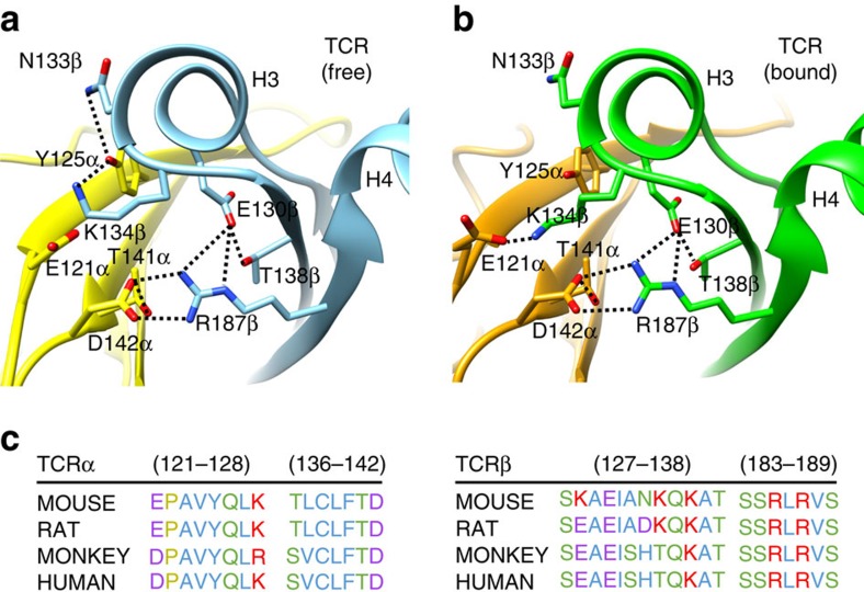
Figure 8. Sequence conservation at the Cα- and Cβ-domain interface near the H3 helix of the TCR.
(a) Free and (b) bound X-ray structures of the B4.2.3 αβ TCR dimer showing a network of electrostatic interactions (dashed lines) at the interface between the α- and β-subunit constant domains. The Cα and Cβ domains are shown with yellow/beige and cyan/green in the free/bound structures, respectively. Functionally important residues identified by NMR to show significant changes upon MHC binding are highlighted on the structure. (c) Sequence conservation patterns within the same regions of the Cα and Cβ domains as in a,b.

