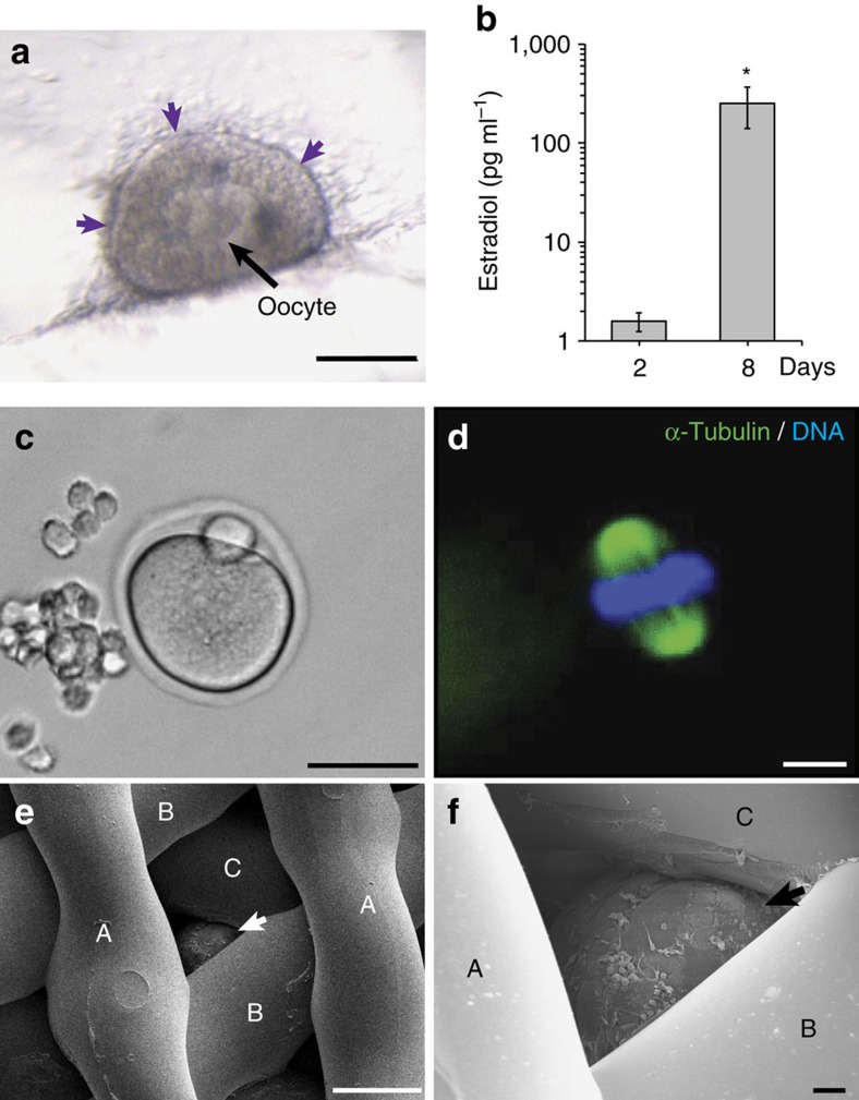
Figure 4. Follicles function within 3D printed scaffolds in vitro.
(a) Light microscopy image of 3βHSD expression (purple) along edge of follicle cultured in 30° scaffold after 4 days of culture. (b) Estradiol secretion in media of follicles cultured in 3D printed scaffolds collected at day 2 and day 8 of culture. (c,d) MII egg with extruded polar body was released from a follicle cultured in 60° scaffold and contains condensed chromatin (blue) along the spindle (green). (e,f) Scanning electron micrograph demonstrating follicle (arrows) wedged underneath three layers of 60° scaffold struts (identified as layers A, B, C) and cultured for 2 days. Scale bars: (c) 50 μm; (a,e) 100 μm; (d,f) 10 μm.

