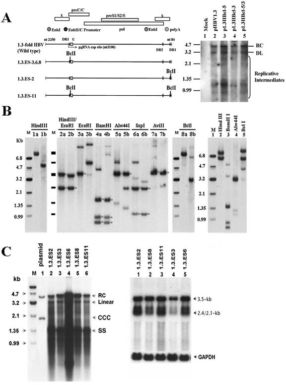
FIG. 3.
Characterization of stable HBV transfectants bearing the BclI marker(s) within a terminal repeated region(s). (A) (Left) Schematic representation of modified 1.3-fold HBV plasmids and the position(s) of their BclI genetic marker(s). (Right) The replication capacity of each BclI-modified plasmid (p1.3HBcl-5, p1.3HBcl-3, and p1.3HBcl-5/3) was examined and compared with that of wild-type HBV. Three days after transient transfection, the viral replicative intermediates were extracted for Southern blot analysis. (B) (Left and center) The chromosome-integrated features of 1.3.ES2 and 1.3.ES11 cells were determined by restriction enzyme digestion. The symbol “a” indicates the restriction enzyme digestion of genomic DNA extracted from 1.3ES2 cells, while “b” indicates genomic DNA prepared from 1.3ES11 cells. Bands marked with asterisks represent the predicted HBV restriction fragments after hybridization with an HBV-specific probe. (Right) Restriction enzyme digestion revealed the chromosome-integrated features of 1.3.ES8 cells. (C) (Left) Cytoplasmic viral replicative intermediates of BclI-bearing HBV lines were extracted to analyze their viral replication ability by Southern blotting. (Right) Total RNAs of various lines were extracted, and viral transcript expression profiles were determined by Northern blotting after probing with an HBV-specific probe. The expression of GAPDH was monitored as a loading control.

