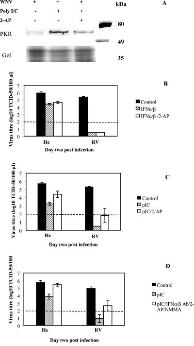
FIG. 4.
PKR contribution to antiviral responses against WN virus upon priming with either IFN-α/β or pIC in primary mouse macrophages from susceptible and resistant mice. (A) Western blot analysis of PKR activity. The levels of phosphorylated PKR were analyzed by Western blot in macrophage lysates obtained from susceptible mice. Cells were isolated and cultivated on 6-well tissue culture plates as described in Materials and Methods. Priming with 1 μg of pIC/ml was performed for 1 h at 37°C in the absence or presence of 2 mM 2-AP. Control cells did not have any treatment. Twenty-four hours later, the cells were lysed in 100 μl of Laemmli sample buffer, and equal amounts were loaded onto SDS-10% PAGE gel in parallel with protein standards (Prestained Broad Range; Bio-Rad). The molecular weights in kilodaltons of the protein standards bovine serum albumin (80 kDa), ovalbumin (49 kDa), and carbonic anhydrase (35 kDa) are shown on the right. The amount of cellular proteins applied was additionally estimated by Coomassie blue staining of the gel below the ovalbumin protein standard. Monoclonal rabbit anti-phospho-PKR (pThr451) antibody (Sigma) was used as a primary antibody, while HRP-conjugated goat anti-rabbit IgG was used as a secondary antibody (Zymed Laboratory). (B and C) Effects of 2-AP inhibitor on IFN-α/β-induced (B) and pIC-induced (C) antiviral responses were determined on day 2 p.i. Average virus titer values were derived from two experiments with four replicas. Student's t test indicated significant differences in virus titers between control samples and each of the treatments (P < 0.001), while no significant difference was estimatedbetween other treatments (P > 0.1). (D) Effect of IFN-α/β Ab in the absence or presence of 2-AP and NMMA inhibitors on the antiviral responses induced by pIC was determined on day 2 p.i. Average values are derived from three experiments and a minimum of six replicas per treatment. Standard error bars are included where appropriate. Student's t test indicated significant difference between virus titers in control infections and those derived from pIC-primed cells for both susceptible and resistant cell cultures (0.001 < P < 0.01 and P < 0.001, respectively), while simultaneous treatments with IFN-α/β Ab, 2-AP, and NMMA did not restore pIC-inhibited WN virus replication back to the control values in cells from resistant mice (P = 0.02). A dashed line denotes a threshold value of 2.0 log10 TCID50 units for accurate detection of virus titers, while virus present below the threshold value was detected as described in the legend for Fig. 1A.

