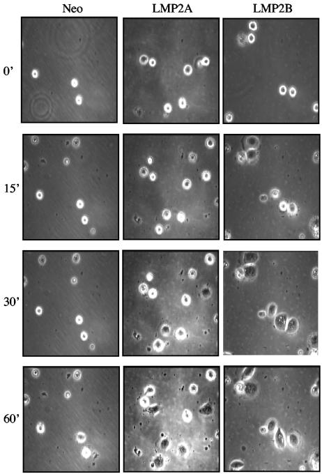
FIG. 3.
Epithelial cells expressing LMP2A and LMP2B show increased rates of attachment and spreading on extracellular matrix. Neo control and LMP2A- and LMP2B-expressing HaCat epithelial cells were serum starved, collected as single cells, and plated onto fibronectin-coated petri dishes. Time-lapse video microscopy was used to analyze the kinetics of cell attachment and spreading over a 60-min time frame, with frames taken every 15 min. Magnification, ×100. Serum starvation and suspension culture resulted in a loss of cell viability that was 18% (±1.7%) in Neo control cells, 31% (±2.6%) in LMP2A-expressing cells, and 29% (±1.5%) in LMP2B-expressing cells (data are from three independent experiments).

