FIG. 6.
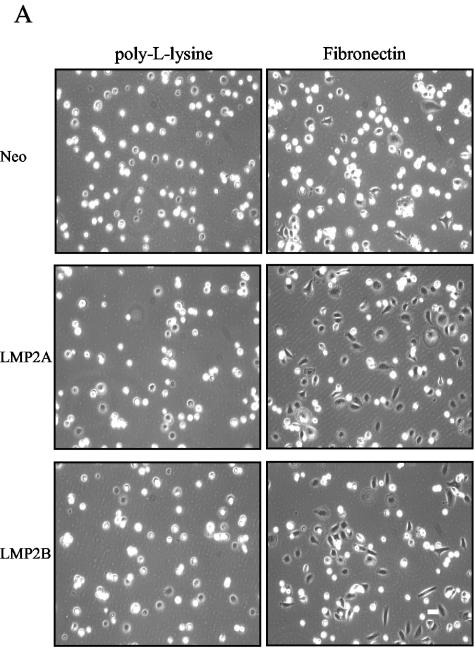
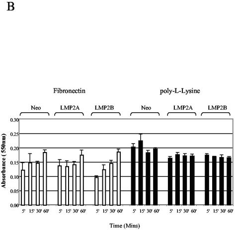
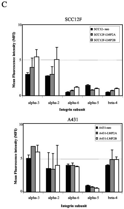
LMP2A and LMP2B promote cell spreading rather than attachment on extracellular matrix. (A) Neo control and LMP2A- and LMP2B-expressing SCC12F cells were serum starved, collected as single cells, and plated onto petri dishes coated with fibronectin or poly-l-lysine. The extent of cell attachment and spreading was visualized after 60 min by phase-contrast microscopy. Magnification, ×50. (B) Attachment of Neo control and LMP2A- and LMP2B-expressing cells to extracellular matrix (fibronectin and poly-l-lysine) are similar, indicating that LMP2A and LMP2B promote cell spreading rather than attachment. Serum-starved cells were plated onto 96-well plates precoated with fibronectin or poly-l-lysine, and the number of adherent cells were quantitated at various times after plating. Data are presented as raw values (OD550) from triplicate determinations (one-way analysis of variance showed no significant differences in the rates of adhesion between Neo control cells, LMP2A-expressing cells, and LMP2B-expressing cells at the time points indicated). (C) The integrin profiles of Neo control and LMP2A- and LMP2B-expressing SCC12F and A431 cells were analyzed by cytofluorimetric (FACS) analysis using a panel of MAbs specific for the α2, α3, α5, α6, and β4 integrin subunits. Data are presented as mean fluorescence intensity (MFI) on an arbitrary scale. Data shown are the means ± standard deviations from three independent experiments.


