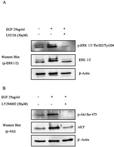
FIG. 9.
Validation of pharmacological inhibitors. SCC12F cells were serum starved in the presence or absence of various pharmacological inhibitors for 30 min prior to stimulation with EGF (25 ng/ml). The ability of 30 μM U0126, 50 μM LY294002, and 5 to 50 μM PP2 to block EGF-induced ERK, Akt, and tyrosine phosphorylation was then analyzed by Western blotting using antibodies specific for phosphorylated forms of ERK and Akt and phosphotyrosine. The ability of 30 μM calphostin C to block PKC activation was assessed in SCC12F cells treated for 30 min with 100 ng of TPA/ml. PKC activation was assessed by determining the extent of PKCα translocation in response o TPA stimulation. Arrows denote plasma membrane-associated PKCα. Bar, 10 μm.

