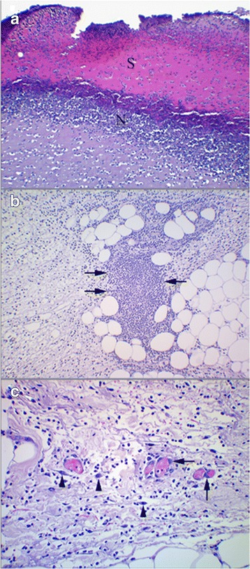
Fig. 4.

Wounds day 2. a: Blue Iris mink. A scab of non-vital tissue (S) demarcated by a zone of neutrophils (N). HE. ×100. b: Silverblue mink. Micro-abscess (arrows) located in edematous subcutis beneath the wound surface. HE. ×100. c: Silverblue mink. Blood filled vessels showing plumping of endothelial cells (arrows). Fibroblasts (arrow heads) in various stages of activation can be seen. HE. ×200
