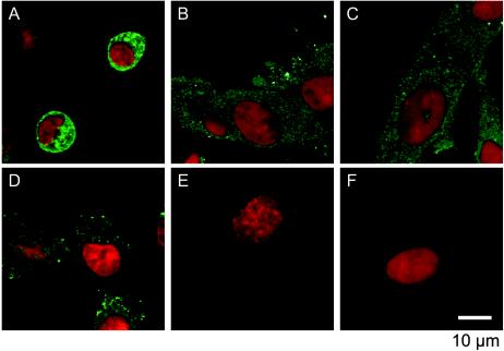
FIG. 3.
PV antigens in PV-infected neural cells with or without MAbs. PV-infected cells were examined in an immunofluorescence study 11 hpi (A, B, and C) and 24 hpi (D, E, and F). At 2 hpi the cells were not treated with MAb (A and D) or were treated with MAb against PV (B and E) or against hPVR (C and F). Red indicates nucleic acids, and green indicates PV antigens.

