
Fig. 1.
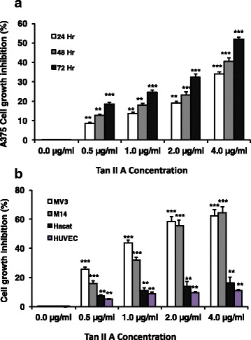
Tan II A inhibited A375, MV3, M14, Hacat, and HUVEC cells proliferation in a dose and time dependent manner. A375 cells were treated with Tan IIA (0.5, 1, 2, or 4 μg/mL) for 24, 48, or 72 h respectively. MV3, M14, Hacat (spontaneously transformed aneuploid immortal keratinocyte cell line from adult human skin), and human umbilical vein endothelial cells (HUVEC) cells were treated with Tan IIA (0.5, 1, 2, or 4 μg/mL) for 72 h respectively. DMEM 10% FBS with DMSO (0.1%) (0.0 μg/mL Tan II A) was used as the negative control. The MTT reading level in the negative control cell was used as 0% cell inhibition. The MTT reduction amount in Tan II A treated cells compared to the negative control was normalized with the negative control (0.1% DMSO) MTT level to calculate the inhibition percentage. *P < 0.05, ** P < 0.01, and ***P < 0.001 for comparison of indicated treatments to the negative control at 24, 48, or 72 h respectively. n = 6 for treatment. a. Tan IIA treatment significantly increased the inhibition of A375 cells proliferation at 24, 48, and 72 h in 0.5, 1, 2, or 4 μg/mL Tan II A compared to the respectively negative controls. b. Tan IIA treatment significantly enhanced the inhibition of MV3, M14, Hacat, and HUVEC cells proliferation at 72 h in 0.5, 1, 2, or 4 μg/mL Tan II A compared to the respectively negative controls