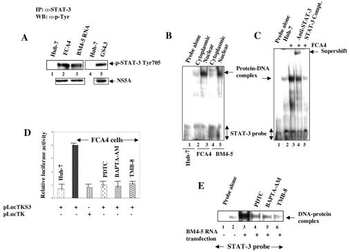
FIG. 1.
HCV replicons induce tyrosine phosphorylation of STAT-3. (A) Whole-cell lysates from cells expressing HCV replicons were immunoprecipitated with anti-STAT-3 serum, fractionated by SDS-PAGE, and immunoblotted with antiphosphotyrosine monoclonal antibody. Lanes 1 and 4, untransfected Huh-7 lysates; lanes 2 and 5, FCA4 and GS4.3 cells expressing HCV subgenomic replicons; lane 3, Huh-7 cells transfected with in vitro-synthesized BM4-5 RNA. The bottom panel represents the expression of HCV NS5A in HCV replicon-expressing cells. (B) An EMSA was carried out in the presence of γ-32P-labeled STAT-3 cognate nucleotide probe, and the nuclear lysates were prepared from FCA4 cells and Huh-7 cells transiently transfected with in vitro-synthesized BM4-5 RNA. Lane 1, STAT-3 probe incubated with Huh-7 nuclear lysates; lanes 2 and 3, equal amounts of FCA4 cytoplasmic and nuclear lysates, respectively; lanes 4 and 5, equal amounts of BM4-5 RNA-transfected cytoplasmic and nuclear lysates, respectively. (C) EMSA. Lane 1, probe alone; lanes 2 and 3, equal amounts of Huh-7 and FCA4 nuclear lysates; lane 4, DNA-protein complex incubated with anti-STAT-3 serum; lane 5, DNA-protein complex treated with a 100-fold excess of unlabeled STAT-3 oligonucleotide. (D) Luciferase reporter gene assay. FCA4 cells were transfected with STAT-3-responsive pLucTKS3 and pLucTK (without STAT-3 binding sites) luciferase plasmids. At 36 h posttransfection, cells were treated with PDTC (6 h), BAPTA-AM (2 h), and TMB-8 (4 h) at various times before preparing the lysates for luciferase activity. (E) EMSA. Lane 1, probe alone; lanes 2 and 3, equal amounts of untransfected and BM4-5 replicon RNA-transfected nuclear lysates; lanes 4, 5, and 6, BM4-5 RNA-transfected lysates treated with PDTC (6 h), BAPTA-AM (2 h), and TMB-8 (4 h) at various times.

