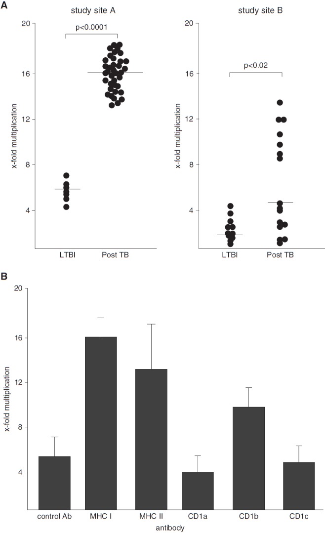
Figure 1.

Effect of bronchoalveolar lavage (BAL) cells on the growth of Mycobacterium tuberculosis (Mtb). A total of 2 × 10⁵ freshly isolated BAL cells were incubated with 1 × 10⁶ Mtb cells. The number of colony-forming units was determined 2 hours and 5 days after infection by plating cell lysates. (A) The x-fold multiplication between 2 hours and 5 days of all donors tested is shown. Experiments were performed at different study sites (Ulm [left panel] and Borstel [right panel]). P values were calculated using two-tailed Mann–Whitney U tests. Horizontal bars represent the medians. (B) A total of 2 × 10⁵ BAL cells from donors with latent tuberculosis infection (LTBI) were preincubated with neutralizing antibodies (10 μg/ml) against antigen-presenting molecules for 1 hour before 1 × 10⁶ Mtb cells were added. The number of colony-forming units was determined after 2 hours and 5 days by plating cell lysates. The figure presents the average x-fold multiplication ± SEM from three different donors. Ab = antibody; MHC = major histocompatibility complex; TB = tuberculosis.
