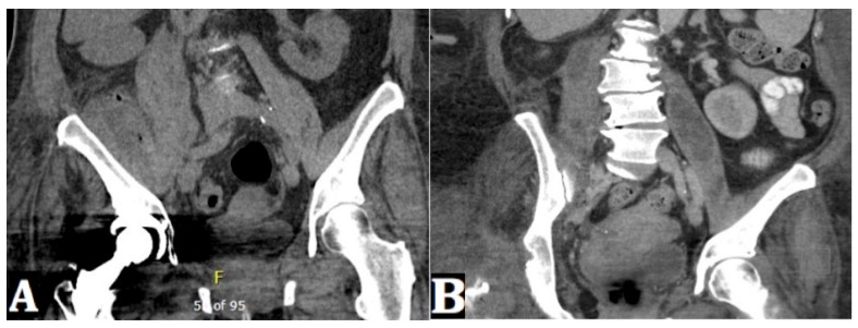
Figure 1.
(A) A coronal CT pelvis image shows fluid and gas within the enlarged right iliacus and iliopsoas musculature. (B) A repeat coronal CT pelvis image (at one month from index imaging) shows an interval development of fluid collection within the right posterior pararenal space/right psoas muscle while there has been interval resolution of loculated fluid collection within the right iliacus muscle region. Right hip hardware has been removed. Additionally, there has been interval development of a loculated fluid collection within the left psoas muscle.

