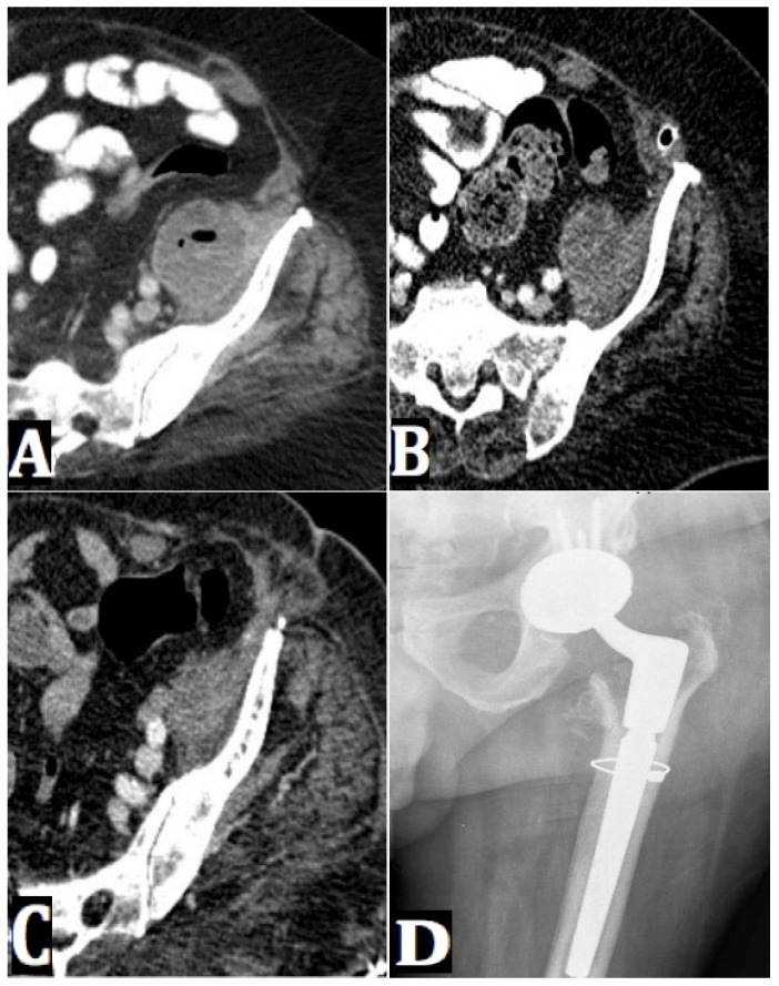
Figure 2.
(A) An axial CT pelvis image shows a fluid collection in the left iliacus muscle. (B) A repeat axial CT pelvis image (at six weeks from index imaging) shows no significant change in size of the left iliacus abscess. (C) A repeat axial CT pelvis image (at four months from index imaging) shows a decrease in size of the left iliacus abscess. This occurred following three retroperitoneal debridements, and two spacer exchanges. (D) An AP radiograph of the left hip shows a left hip revision arthroplasty with a cerclage wire in proximal femur and components in expected position. There is no evidence of radiographic loosening at 10 months post-op.

