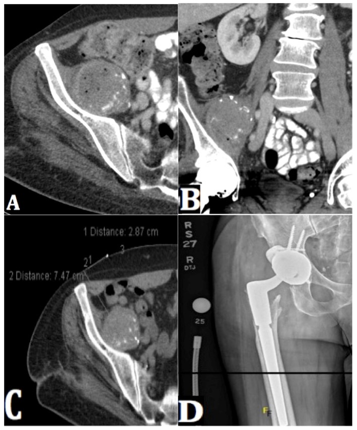
Figure 3.
(A) An axial CT pelvis shows fluid distension with intermittent gas in the right iliacus muscle, also containing scattered calcifications. (B) A coronal CT pelvis image shows fluid distension with intermittent gas in the right iliacus muscle, also containing scattered calcifications. This was biopsy and aspirate proven to be consistent with infected hematoma. Notably, the patient was on Coumadin following his initial primary THA operation. (C) A repeat axial CT pelvis image reveals a persistent right iliacus abscess. (D) An AP radiograph of the right hip shows a right hip revision arthroplasty with hardware intact and no evidence of loosening at four months post-op.

