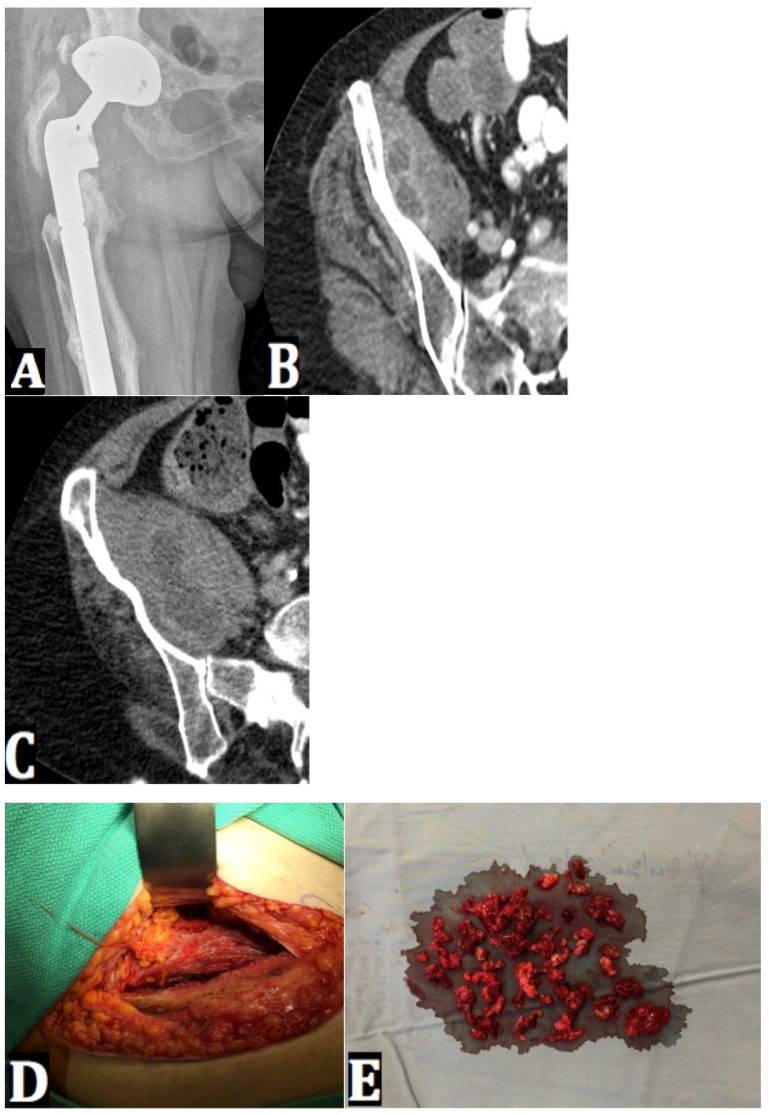
Figure 4.
(A) An AP radiograph of the right hip shows a revision hip prosthesis with minimal femoral bone stock and lucency surrounding the femoral stem. The component position has not changed over a ten-year period. (B) An axial CT right hip image shows new enlargement of right iliacus muscle containing multi-loculated fluid collection. (C) A repeat axial CT right hip image of the right hip shows an interval increase in size of the collection in the right iliacus muscle. (D) An intraoperative photo of the retroperitoneal approach to the inner table of pelvis shows the point of access to the proximal belly of the iliacus muscle. (E) An intraoperative photo shows debrided iliacus muscle abscess tissue.

