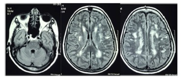
Figure 1. Brain magnetic resonance imaging (MRI, axial flair) showing hyper intense multiple lesions in the periventricular and subcortical white matter in a 56-year-old female HAM/TSP patient with 10 years of disease symptoms and motor disability grade 4.

