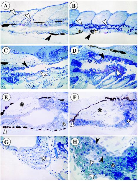
FIG. 4.
Histopathology of zebrafish embryos infected with SHRV by immersion. (A) Perianal region of control fish. White arrowheads indicate normal blood vessels. The black arrowhead indicates a normal pigment cell. Magnification, ×400. (B) Perianal region of infected fish. The white arrowheads indicate a blood vessel filled with monocytes. The black arrowhead indicates an irregularly shaped pigment cell. Magnification, ×400. (C) Branchial region of control fish, with normal mucus cells of the branchial epithelia (white arrowhead). Magnification, ×400. The black arrowhead points to the upper pharyngeal epithelium. (D) Branchial region of infected fish. The upper pharyngeal epithelium has a rough structure and contains many proliferating cells (black arrowhead). The white arrowhead indicates numerous pink mucus cells. Magnification, ×400. (E) Liver (white asterisk) and swim bladder (black asterisk) of control fish. The white arrowhead indicates normal pigment cells. Magnification, ×250. (F) Dark staining liver (white asterisk) and congested swim bladder (black asterisk) of infected fish. The white arrowhead indicates irregularly shaped pigment cells of infected fish. magnification, ×250. (G) Higher magnification of liver tissue (white asterisk) from control fish. Magnification, ×400. (H) Higher magnification of liver tissue (white asterisk) from infected fish. Black arrowheads indicate intracellular vacuoles. White arrowheads indicate glycogen vesicles in the extracellular space. Magnification, ×1000.

