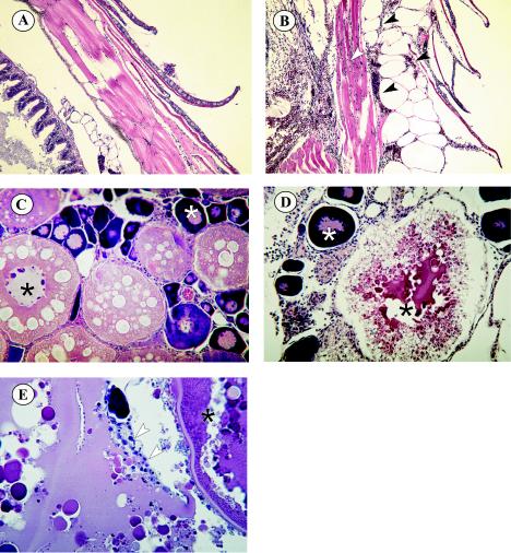
FIG. 5.
Histopathology of adult zebra fish infected with SHRV by i.p. injection. (A) Normal scales and epidermis of control fish. Magnification, ×100. (B) Scales and epidermis of infected fish. Black arrowheads indicate subdermal edema and hemorrhaging. The white arrowhead indicates hemorrhaging in the underlying muscle tissue. Magnification, ×100. (C) Ovaries of control fish showing normal egg development, with generations of ova in different developmental stages. The white asterisk indicates a primary oocyte, the black asterisk indicates a secondary oocyte, and the black arrowhead indicates the epithelial granulosa (nursing) cells. Magnification, ×200. (D) Degenerating secondary oocyte of SHRV-infected fish (black asterisk). Primary oocytes seem to be unaffected (white asterisk). Magnification, ×200. (E) Epithelial granulosa cells (white arrows) reabsorbing remaining yolk from secondary oocyte.

