
Figure 2. Region based gray matter volume analysis in major depressive disorder using cytoarchitectonic maps of the human frontal pole.
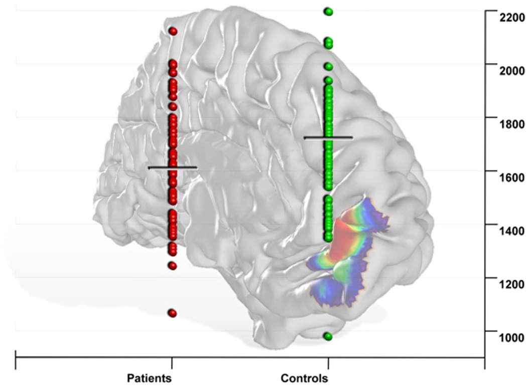
Significant decrease of volume only for the left medial part of the frontal pole (left area Fp2) in depressed patients compared to controls (p<0.05; FDR corrected).