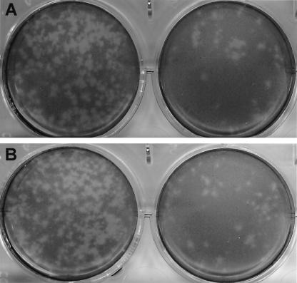
FIG. 5.
Plaque-forming assay of pCV4A viruses measured at dilutions of 10−4 (left cultures) and 10−5 (right cultures) in the presence of IC (1%) (A) or GCDCA (200 μM) (B). After LLC-PK cells were infected for 1 h with the viruses recovered from pCV4A, the cells were placed under medium containing MEM, agarose (1%), neutral red (0.002%), and IC (1%) or GCDCA (200 μM). Pictures were taken 96 h after virus inoculation.

