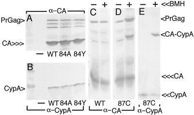
FIG. 5.
CypA assembly into virus particles. (A and B) 293T cells were mock transfected (−) or transfected with the indicated constructs, and medium supernatants were collected 72 h posttransfection and then subjected to ultracentrifugation. Pelleted virus samples were resuspended in buffer and subjected to SDS-PAGE and parallel immunodetection with the indicated antibodies. (C to E) Samples were mock treated (−) or treated (+) with the cysteine-specific cross-linker BMH prior to SDS-PAGE and immunodetection. CypA, CA, PrGag, and CA-CypA cross-link products (D and E, cross-link lanes) are indicated. Note a putative CA-CA dimer cross-link product in the panel D cross-link lane, just above the PrGag band.

