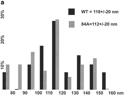
FIG. 8.
Analysis of virus morphologies. Virus particles from wt HIVLuc- and H84A HIVLuc-transfected cells were isolated, lifted onto carbon-coated grids, stained with uranyl acetate, and imaged by transmission EM. (a) Histogram of particle diameters. Diameters of wt (n = 29) and H84A (n = 39) virus particles were plotted in 10-nm size bins relative to the frequencies (percentage of total sample) observed for each size bin. (b) Galleries of virus particle images. Images of eight wt (A to H) and H84A (I to P) virus particles are shown within 262- by 262-nm windows. Note that panel H shows a wt core, apparently released from a broken virus during preparation. Note also that all wt virions (n = 29) showed discernible cylindrical or conical cores, whereas only panel I showed a possible cylindrical core for H84A (n = 39).


